| x | x | ||||
|
|
 |
||||
| BACTERIOLOGY | IMMUNOLOGY | MYCOLOGY | PARASITOLOGY | VIROLOGY | |
|
|
|
||||
| SPANISH | |||||
| PORTUGUESE | |||||
| ALBANIAN | |||||
|
Let us know what you think FEEDBACK |
|||||
| SEARCH | |||||
|
|
|||||
|
|
|||||
|
TEACHING OBJECTIVES An overall view of the replication cycle of viruses |
PRINCIPAL EVENTS INVOLVED IN REPLICATION
Uncoating Synthesis of viral nucleic
acid and protein Assembly/maturation Release
STRUCTURAL VERSUS NON-STRUCTURAL PROTEINS All proteins in a mature virus particle are said to be structural proteins - even if they make no contribution to the morphology or rigidity of the virion - non-structural proteins are those viral proteins found in the cell but not packaged into the virion. |
||||
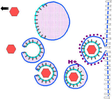
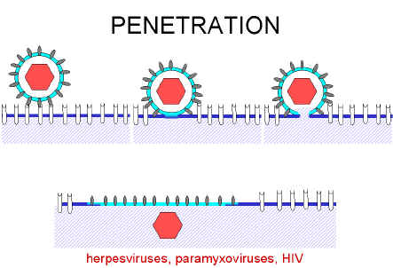 Figure 1. Fusion of a virus with the plasma membrane after
attachment to a cell surface receptor
Figure 1. Fusion of a virus with the plasma membrane after
attachment to a cell surface receptor
|
|||||
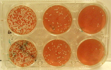 Figure 5.
A plaque assay. Serial dilutions of virus have been plated on confluent monolayer cultures of cells. The cells are stained after a period of time in which a single virus infects a cell, produces new virus particles and infects surrounding cells. The white areas show areas of the culture in which the cells have been killed. Each "plaque" is the result of the presence of one original infectious virus particle.
|
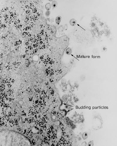
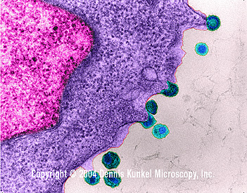
EFFECT OF VIRUSES ON HOST MACROMOLECULAR SYNTHESIS Many viruses inhibit host RNA, DNA or protein synthesis (or any combination of these). The mechanisms by which the virus does this vary widely. Cytopathic effect (CPE) The presence of the virus often gives rise to morphological changes in the host cell. Any detectable changes in the host cell due to infection are known as a cytopathic effect. Cytopathic effects (CPE) may consist of cell rounding, disorientation, swelling or shrinking, death, detachment from the surface, etc. Many viruses induce apoptosis (programmed cell death) in infected cells. This can be an important part of the host cell defense against a virus - cell death before the completion of the viral replication cycle may limit the number of progeny and the spread of infection. (Some viruses delay or prevent apoptosis - thus giving themselves a chance to replicate more virions.) Some viruses affect the regulation of expression of the host cell genes which this can have important results both for the virus's ability to grow, and in terms of the effect on the host cell. The cytopathic effects produced by different viruses depend on the virus and the cells on which it is grown. This can be used in the clinical virology laboratory to aid in identification of a virus isolate.
The CPE effect can be used to quantitate infectious virus particles by the plaque-forming unit assay (figure 5). Cells are grown on a flat surface until they form a monolayer of cells covering a plastic bottle or dish. They are then infected with the virus. The liquid growth medium is replaced with a semi-solid one so that any virus particles produced as the result of an infection cannot move far from the site of their production. A plaque is produced when a virus particle infects a cell, replicates, and then kills that cell. Surrounding cells are infected by the newly replicated virus and they too are killed. This process may repeat several times. The cells are then stained with a dye which stains only living cells. The dead cells in the plaque do not stain and appear as unstained areas on a colored background. Each plaque is the result of infection of one cell by one virus followed by replication and spreading of that virus. However, viruses that do not kill cells may not produce plaques. Other assays for viruses
|
||||
|
|
|||||
|
WEB RESOURCES
|
|
||||
|
|
|||||