![]()

VIROLOGY
Appendix
EBOLA VIRUS
Dr Richard Hunt
Professor
University of South Carolina School of Medicine
FEEDBACK
 Figure 1
2014 Ebola Outbreak in West Africa - Outbreak Distribution Map
CDC
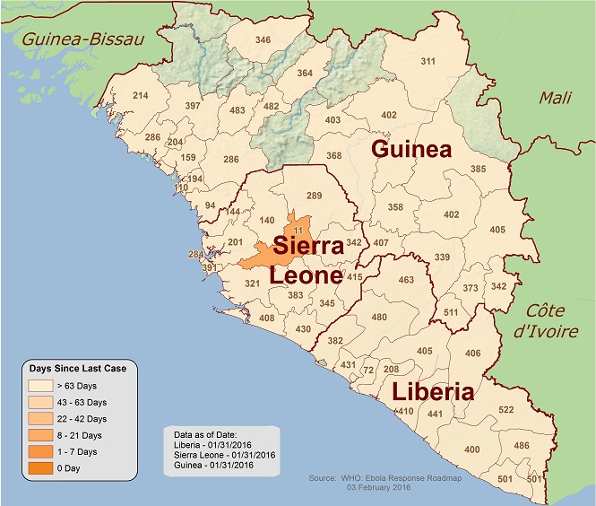 Figure 2
2014 Ebola Outbreak in West Africa - Days since last case. Situation as of
January 31, 2016
CDC
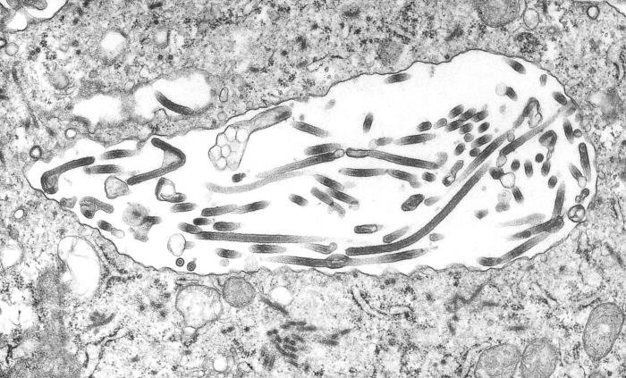 Figure 3
Ultrastructural morphologic changes in a tissue sample from a patient with
an Ebola hemorrhagic fever infection, including the presence of numbers of
Ebola virions.
CDC - Cynthia Goldsmith
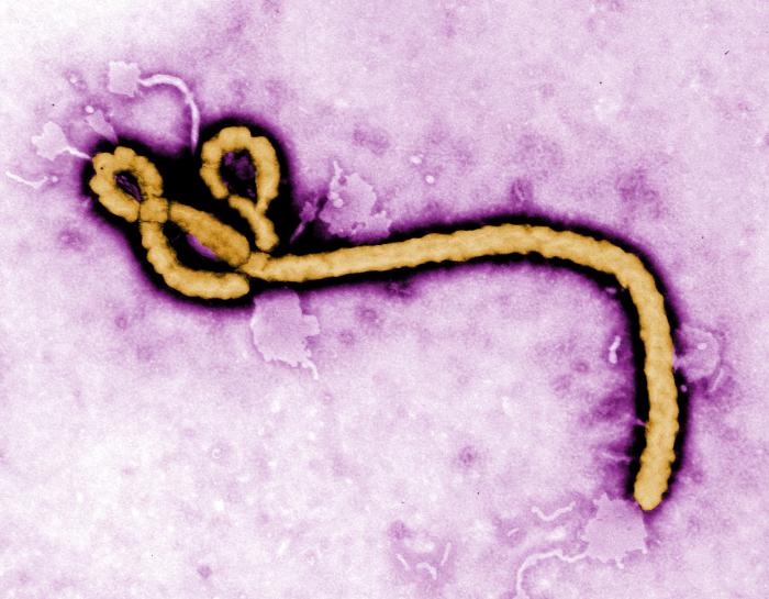 Figure 4
Colorized transmission electron micrograph showing the ultrastructural
morphology displayed by an Ebola virus virion
CDC/ Frederick A. Murphy
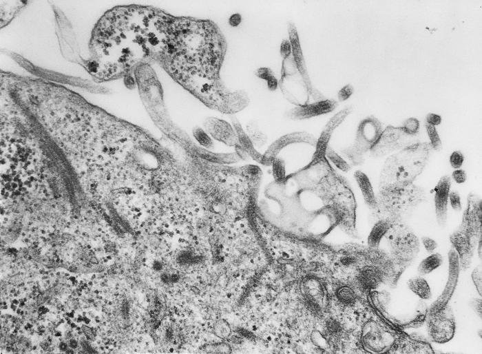 Figure 5
Thin section electron micrograph of cell shedding Ebola virus
CDC
 Figure 6
Colorized Transmission Electron Micrograph of the Ebola Virus.
CDC - Cynthia Goldsmith
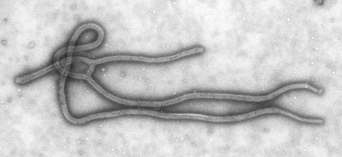 Figure 7
Ebola Virus
CDC - Cynthia Goldsmith
Ebola virus causes Ebola hemorrhagic fever (EHF), a severe disease (the fatality rate is 55 - 60%) that occurs in humans and some other primates including gorillas and chimpanzees.
It was first thought that humans contracted disease as a result of eating gorilla or chimpanzee meat but that is now thought to be unlikely. EHF is a zoonotic disease and although the reservoir host is not know precisely, much evidence points (as in several other important zoonotic diseases) to bats. Specifically, contact with bat urine, saliva or feces.
The virus was first associated with human disease in 1976 when an outbreak occurred in the Democratic Republic of the Congo (DRC) near the Ebola River in which there were 602 reported cases of hemorrhagic fever and 431 patients died. There have been several more recent outbreaks including in 1995 also in the DRC (315 cases and 254 deaths), in 2000 in Uganda (425 cases and 224 deaths), and in 2007 in Uganda and the DRC again (413 cases and 214 deaths).
In 2014, the largest Ebola outbreak so far occurred. By April 2016, the number of suspected and confirmed cases was 28,652 with 15,261 laboratory confirmed cases and 11,325 deaths (figs. 1 and 2). This epidemic is thought to have started in a village in Guinea in an area in which people often hunt bats.
Ebola Virus
Ebola virus (figs. 3 - 7) is a member of the Filovirus family (Filoviridae) which also contains Marburg virus (a human pathogen, discovered in 1967 in Marburg, Germany causing Marburg hemorrhagic fever) and Cuevavirus (discovered in 2010 in bats in Europe). Filoviruses have a long filamentous shape. They are about 80nm in diameter and hundreds of nanometers long. The longest are about 14 µm long. They are enveloped, acquiring their membrane from the plasma membrane of the host cell, and bear a glycosylated fusion protein on their surface which gives them a spiked appearance. The genome is single-stranded negative sense RNA (about 19kB with seven genes) and the viruses replicate exclusively in the cytoplasm of the infected cell from which they bud through the plasma membrane.
There are five known Ebola virus species:
Ebola virus (formerly named Zaire Ebola virus). This is the most lethal of the Ebolaviruses.
Bundibugyo Ebolavirus
Sudan Ebolavirus
Tai Forest Ebolavirus
Reston Ebolavirus. This was first discovered in crab-eating macaques in a laboratory at Reston, Virginia in the United States and appears not to be pathogenic to humans.
EBOLA HEMORRHAGIC FEVER
Infection
Outbreaks of EHF are sporadic and exactly how humans pick up the virus from the reservoir host is unknown. Indeed, though highly suspected to be bats, the reservoir host has yet to be unequivocally identified. It is probable that humans acquire the virus from saliva, feces and urine of infected bats such as by eating contaminated food. The virus is usually not transmitted in aerosols (as is the case with flu) but is spread by direct, close personal contact with the bodily fluids (blood, urine, sweat) of an infected person. Thus, if the infected patient is isolated and contact personnel wear protective clothing, the outbreak should be contained. Most common transmission is to persons who care for a patient (such as washing) or other close contact. In areas where outbreaks occur, the hospital facilities are often unsanitary and nosocomial infections occur as a result of direct contact with bodily fluids, unsterilized needles and medical equipment.
It has been found that even after an apparent recovery, the virus may remain in some body fluids, including semen. It appears that the rate of elimination of Ebola from the semen is different for each man. It may last for three to nine months. It appears that the amount of virus decreases over time.
Sexual transmission
Classically, Ebola virus is transmitted via body fluids such as urine and blood of an infected person. In 2015, it was shown that men who have survived the disease can have the virus in their semen. The testis is an immune-privileged site and viruses that might be cleared from elsewhere in the body may persist here. Viral RNA could be detected by RT-PCR for up to 9 months from the start of the infection. In the current 2014 outbreak, there are estimated to be about 8,000 male survivors but there have been only about 20 suspected cases of sexual Ebola virus transmission.
Symptoms
The Centers for Disease Control (CDC) list the following symptoms for EHF:
Fever
Headache
Joint and muscle aches
Weakness
Diarrhea
Vomitting
Stomach pain
Lack of appetite
Some patients may experience:
Rash
Red eyes
Hiccups
Cough
Sore throat
Chest pain
Difficulty breathing
Difficulty swallowing
Bleeding inside and outside the body
Symptoms may appear anywhere from 2 to 21 days after exposure to the virus although 8-10 days is most common. Some who become sick with EHF are able to recover, while others do not. The reasons behind this are not yet fully understood. However, it is known that patients who die usually have not developed a significant immune response to the virus at the time of death.
In about half of cases, the disease progresses to EHF. Internal hemorrhage leads to blood in vomit, urine and feces. It can also be seen under the skin and around the eyes and mouth. Unseen bleeding also occurs internally and this loss of fluid causes a dramatic drop in blood pressure leading to organ failure. It is the latter that kills the patient.
Countries in which EHF has occurred
In Africa, confirmed cases of Ebola HF have been reported in (CDC):
Guinea
Liberia
Sierra Leone
Nigeria
Democratic Republic of the Congo (DRC)
Gabon
South Sudan
Ivory Coast
Uganda
Republic of the Congo (ROC)
South Africa (imported)
Diagnosis
Initial diagnosis is difficult because of the non-specific nature of the symptoms. If the initial stages of EHF are suspected (such as recent travel in an Ebola virus-infected area), there are specific tests available. ELIZA tests for specific Ebola proteins, PCR and direct isolation of the virus may be used. Later the patient develops IgM and IgG antibodies.
Supportive Treatment
In most situations, treatment is palliative. The patient’s electrolytes and body fluids, oxygen levels and blood pressure are normalized. Any other infections that the patient has are also treated.
Antibody treatment - ZMapp
There is a more specific experimental treatment that has been tried with two Americans who were infected in Africa in the 2014 outbreak and returned to Emory University Hospital in the United States for further care. This therapy is now being made available in West Africa. The treatment is called ZMapp (because it is made by Mapp Biopharmaceuticals). It is not a vaccine because vaccines consist of antigens from the pathogen that stimulate the host’s own immune response. Rather, ZMapp consists of already formed exogenous antibodies against Ebola virus proteins. Hybridomas are produced from mice that have been injected with Ebola proteins and the genes encoding the anti-Ebola antibodies (the antigen binding site (Fab)) extracted. The genes are then genetically engineered to produce a gene that has much of the mouse antibody protein replaced by human protein. Only the active site, the antigen binding site, remains as a mouse amino acid sequence. This is called humanization of the mouse monoclonal antibody. The engineered gene is then cloned into a plant transformation vector and using a system based on Agrobacterium, Nicotiniana (tobacco) plants are infected. The tobacco plants then produce the humanized antibody. ZMapp contains two monoclonal antibodies (MB003 and ZMab) both of which have been shown to be effective in very small post-exposure trials with rhesus macaques.
Natural Immunity of Ebola
EHF is a very uncommon disease and, though alarming, kills far fewer people than diseases such as malaria. The French Institut de Recherche pour la Developpment (IRD) found that a large number of people in Gabon have anti-Ebola antibodies. In rural communities, IRD found that 15.3% of the population had these antibodies but they had never had EHF or any disease symptoms. Indeed, many Gabonese people in areas that have never experienced an Ebola outbreak have these antibodies. Presumably they have nevertheless come in contact with infected bat excrement. In some villages the prevalence of anti-Ebola antibodies is as high as 33.8%. Further studies have shown that the people expressing anti-Ebola antibodies have a higher number of T8 lymphocytes. They appear to have a specific immune memory to Ebola virus. IRD researchers suspect that these people have had a mild form of Ebola virus infection that did not produce symptoms. It is likely that these people have developed a natural immunity to subsequent Ebola infection. But why they never developed symptoms is a mystery. Clearly the symptoms of even a non-hemorrhagic Ebola infection are severe and likely to have been remembered.
Vaccine
There is no currently approved vaccine against Ebola; however, several are in development, many of which are based on replication-deficient and replication-competent viruses. A trial is underway in West Africa using a vaccine (rVSV-ZEBOV) based in replication competent vesicular stomatitis virus (VSV) which normally infects cows and other animals but is harmless to humans. The recombinant VSV has been engineered to express the surface glycoprotein of Zaire Ebola virus. In December 2016, it was reported that this vaccine is 70 - 100% effective.
Several other Ebola vaccines are being tested.
Return to Viral Diseases transmitted by vertebrates chapter
This page last changed on Saturday, April 15, 2017
Page maintained by Richard Hunt