|
x |
x |
|
 |
 |
|
INFECTIOUS
DISEASE |
BACTERIOLOGY |
IMMUNOLOGY |
MYCOLOGY |
PARASITOLOGY |
VIROLOGY |
|
|
PARASITOLOGY - CHAPTER FIVE
CESTODES (TAPE WORMS)
Dr Gregory Brower
Professor
University of South Carolina School of Medicine
Dr Abdul Ghaffar
Professor Emeritus
University of South Carolina School of Medicine
|
|
|
|
|
Let us know what you think
FEEDBACK |
|
SEARCH |
|
|

 |
|
Logo image © Jeffrey
Nelson, Rush University, Chicago, Illinois and
The MicrobeLibrary |
|
|
|
|
|
TEACHING
OBJECTIVES
Epidemiology,
morbidity and mortality
Morphology of the organism
Life cycle, hosts and
vectors
Disease, symptoms,
pathogenesis and site
Diagnosis
Prevention and control |
Clinically important cestodes
pathogenic to man are Tenia solium (pork tapeworm), T. saginata
(beef tapeworm), Diphyllobothrium lattum (fish or broad tapeworm), Hymenolepis
nana (dwarf tapeworm) and Echinococcus granulosus and E.
multilocularis (hydatid).
Tenia solium
T. saginata (Teniasis)
Epidemiology
These cestodes have a worldwide distribution but incidence is higher in developing
countries. Infection rate is as low as 1 per 1000 in most of North America and as
high as 10% in the third world. Pork tapeworm shows a higher incidence but this
is dependent on
dietary habits.
Morphology
T. saginata can be
up to 4 to 6 meters long and 12 mm broad; it has a pear-shaped head (scolex) with four suckers but
no hooks or neck. It has a long flat body with several hundred segments (proglottids).
Each segment is about 18 x 6 mm with a branched uterus (15-30 branches). The egg
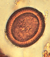
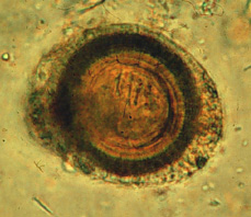
is 35 x 45 micrometers, roundish and yellow-brown. It has peripheral radial striations and contains an embryo with 3
hooklets (figure 2).
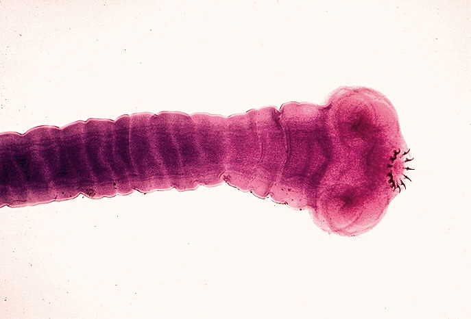
T. solium
is
slightly smaller than T. saginata. It has a globular scolex with four suckers and
a circular row of hooks (rostellum) that gives it a solar appearance. There is a
neck and it has a long
flat body (0.1 meter in length). The proglottids are 5 x 10 mm with a 7-12 branch uterus.
The eggs of T. solium and T. saginata are indistinguishable
(figure 2).
Life cycle
A tapeworm larval cyst
(cysticercus)
is ingested with poorly cooked infected meat; the larva escapes the cyst and
passes to the small intestine where it attaches to the mucosa by the scolex
suckers. The proglottids develop as the worm matures in 3 to 4 months. The adult
may live in the small intestine as long as 25 years and pass gravid proglottids
with the feces. Eggs extruded from the proglottid contaminate and persist on
vegetation for several days and are consumed by cattle or pigs in which they
hatch and form cysticerci (Figure 1).
Symptoms
Light infections remain
asymptomatic, but heavier infections may produce abdominal discomfort,
epigastric pain, vomiting and diarrhea.
Cysticercosis
T. solium eggs
can also infect humans and cause cysticercosis (larval cysts in lung, liver, eye
and brain) resulting in blindness and neurological disorders. The incidence of
cerebral cysticercosis can be as high 1 per 1000 population and may account for up to 20% of
neurological case in some countries (e.g., Mexico); cysticercosis ocular
involvement occurs in about 2.5% of patients and muscular involvement is as high as 10% (India).
Pathology and Immunology
Gastrointestinal symptoms are due to the presence of the tape worm.
Cysticercosis symptoms are a result of inflammatory/immune responses. Antibodies
are produced in cysticercosis and are useful epidemiological tools.
Diagnosis
Diagnosis is based on the
recovery of eggs or proglottids in stool or from the perianal area.
Cysticercosis is confirmed by the presence of antibodies.
Treatment and control
Praziquantel is
the drug of choice. Expulsion of scolex must be assured to assume a satisfactory
treatment. A thorough inspection of beef and pork, adequate cooking or freezing
of meat are effective precautions, since cysticerci do not survive temperatures
below -10o C and above 50o C.
|
|
Figure
1
|
 Figure 1 Figure 1
Life cycle of Taenia saginata and Taenia solium
Humans are the only definitive hosts for Taenia saginata and Taenia
solium. Eggs or gravid proglottids are passed with feces
 ;
the eggs can survive for days to months in the environment. Cattle
(T. saginata) and pigs (T. solium) become infected by
ingesting vegetation contaminated with eggs or gravid proglottids ;
the eggs can survive for days to months in the environment. Cattle
(T. saginata) and pigs (T. solium) become infected by
ingesting vegetation contaminated with eggs or gravid proglottids
 .
In the animal's intestine, the oncospheres hatch .
In the animal's intestine, the oncospheres hatch
 ,
invade the intestinal wall, and migrate to the striated muscles, where
they develop into cysticerci. A cysticercus can survive for
several years in the animal. Humans become infected by ingesting
raw or undercooked infected meat ,
invade the intestinal wall, and migrate to the striated muscles, where
they develop into cysticerci. A cysticercus can survive for
several years in the animal. Humans become infected by ingesting
raw or undercooked infected meat
 .
In the human intestine, the cysticercus develops over 2 months into an
adult tapeworm, which can survive for years. The adult tapeworms
attach to the small intestine by their scolex .
In the human intestine, the cysticercus develops over 2 months into an
adult tapeworm, which can survive for years. The adult tapeworms
attach to the small intestine by their scolex
 and reside in the small intestine
 .
Length of adult worms is usually 5 m or less for T. saginata
(however it may reach up to 25 m) and 2 to 7 m for T. solium.
The adults produce proglottids which mature, become gravid, detach from
the tapeworm, and migrate to the anus or are passed in the stool
(approximately 6 per day). T. saginata adults usually have
1,000 to 2,000 proglottids, while T. solium adults have an
average of 1,000 proglottids. The eggs contained in the gravid
proglottids are released after the proglottids are passed with the
feces. T. saginata may produce up to 100,000 and T.
solium may produce 50,000 eggs per proglottid respectively. CDC DPDx
Parasite Image Library .
Length of adult worms is usually 5 m or less for T. saginata
(however it may reach up to 25 m) and 2 to 7 m for T. solium.
The adults produce proglottids which mature, become gravid, detach from
the tapeworm, and migrate to the anus or are passed in the stool
(approximately 6 per day). T. saginata adults usually have
1,000 to 2,000 proglottids, while T. solium adults have an
average of 1,000 proglottids. The eggs contained in the gravid
proglottids are released after the proglottids are passed with the
feces. T. saginata may produce up to 100,000 and T.
solium may produce 50,000 eggs per proglottid respectively. CDC DPDx
Parasite Image Library

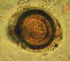 Figure 2A Figure 2A
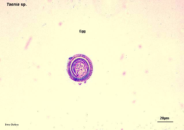
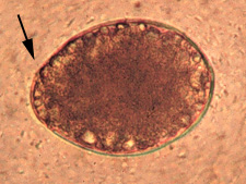
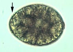
Taeniid eggs. The eggs of Taenia saginata and T. solium are undistinguishable morphologically (morphologic species identification will have to rely on the proglottids or
scolices). The eggs are rounded or subspherical, diameter 31 - 43 µm, with a thick radially striated
brown shell. Inside each shell is an embryonated oncosphere with 6 hooks. The egg in B still has the primary membrane that
surrounds eggs in the proglottids. CDC

 Figure
2B Figure
2B
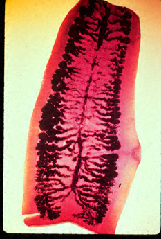
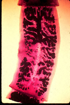
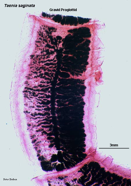
Gravid proglottids of (left) Taenia saginata and (right) T. solium. Injection of India ink in the uterus allows
visualization of the primary lateral branches. Their number allows differentiation between the two species: T. saginata has 15 - 20 branches on each side, while T. solium has 7 - 13. Note the genital pores in mid-lateral
position. CDC
 Figure 2C
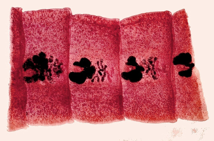
Taenia saginata gravid proglottid
© Dr
Peter Darben, Queensland University of Technology clinical
parasitology collection. Used with permission
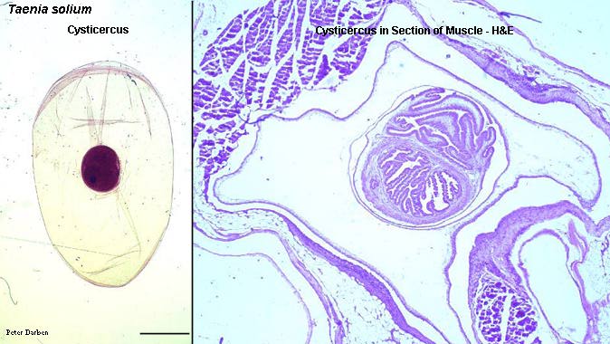
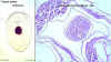 Figure 2D Figure 2D
Taenia solium cysticercus, whole and in section of muscle (H&E)
© Dr
Peter Darben, Queensland University of Technology clinical
parasitology collection. Used with permission
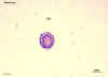 Figure 2E Figure 2E
Taenia sp. egg
© Dr
Peter Darben, Queensland University of Technology clinical
parasitology collection. Used with permission
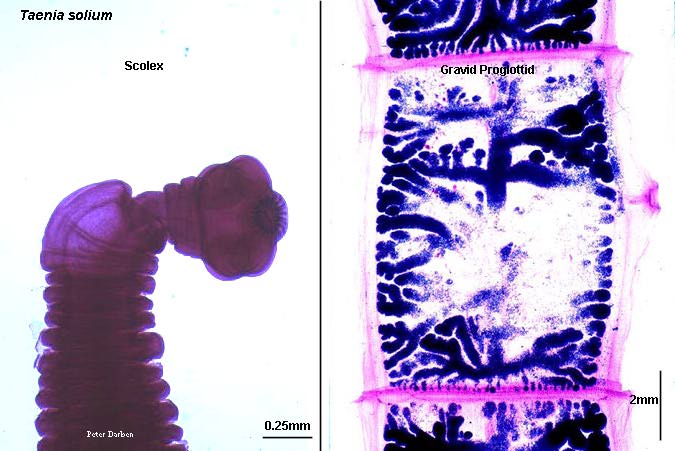
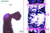 Figure 2F
Taenia solium scolex and gravid proglottid
©
Dr Peter
Darben, Queensland University of Technology clinical parasitology
collection. Used with permission
 Figure 2G
Scolex of Taenia solium.
CDC/Dr. Mae Melvin
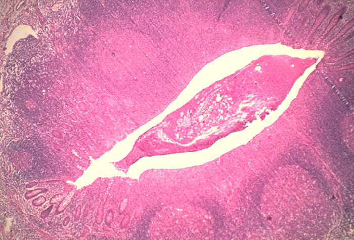
 Figure 2G Figure 2G
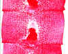
Histopathology of Taenia saginata in appendix. CDC
|
| |
|
|
|
|
|
WEB RESOURCES
Diphyllobothriasis
- CDC |
Diphyllobothrium latum
(fish or broad tapeworm)
Epidemiology
Fish tapeworm infection is
distributed worldwide, in the subarctic and temperate regions; it is associated with
eating of raw or improperly cooked fresh water fish.
Morphology
This is the longest tapeworm found in man, ranging from 3-10 meters with more than 3000 proglottids. The scolex
resembles two almond-shaped leaves and the proglottids are broader than they are
long,
a morphology reflected in the organism's name. Eggs are 30 x 50 micrometers in size and
contain an embryo with 3 pairs of hooklets (figure 4).
Life cycle
Man and other animals are
infected by eating uncooked fish that contains plerocercoid larvae (15 x 2 mm)
which attach to the small intestinal wall and mature into adult worms in 3 to 5 weeks.
Eggs discharged from gravid proglottids in the small intestine are passed in the
feces. The egg hatches in fresh water to produce a ciliated coracidium which
needs to be ingested by a water flea (Cyclops) where it develops into a
procercoid larva. When infected Cyclops are ingested by the freshwater fish, the
procercoid larva penetrates the intestinal wall and develops into a plerocercoid
larva, infectious to man (figure 3).
Symptoms
Clinical symptoms may be
mild, depending on the number of worms. They include abdominal discomfort, loss
of weight, loss of appetite and some malnutrition. Anemia and neurological
problems associated with vitamin B12 deficiency are seen in heavily infected
individuals.
Diagnosis
Diagnosis is based on
finding many typical eggs and empty proglottids in feces (Figure 3). A history of
raw fish consumption and residence in an endemic locality is helpful.
Treatment and control
Praziquantel is
the drug of choice. Freezing for 24 hours, thorough cooking or pickling of fish
kills the larvae. Fish reservoirs should be kept free of raw sewage.
|
|
|
 Figure 3
Immature eggs are passed in feces
.
Under appropriate conditions, the eggs mature (approximately 18 to 20
days) and yield oncospheres
which develop into a coracidia
.
After ingestion by a suitable freshwater crustacean (the copepod
first intermediate host) the coracidia develop into procercoid larvae
 .
Following ingestion of the copepod by a suitable second intermediate
host, typically minnows and other small freshwater fish, the procercoid
larvae are released from the crustacean and migrate into the fish flesh
where they develop into a plerocercoid larvae (sparganum) .
Following ingestion of the copepod by a suitable second intermediate
host, typically minnows and other small freshwater fish, the procercoid
larvae are released from the crustacean and migrate into the fish flesh
where they develop into a plerocercoid larvae (sparganum)
 .
The plerocercoid larvae are the infective stage for humans.
Because humans do not generally eat undercooked minnows and similar
small freshwater fish, these do not represent an important source of
infection. Nevertheless, these small second intermediate hosts can
be eaten by larger predator species, e.g., trout, perch, walleyed pike .
The plerocercoid larvae are the infective stage for humans.
Because humans do not generally eat undercooked minnows and similar
small freshwater fish, these do not represent an important source of
infection. Nevertheless, these small second intermediate hosts can
be eaten by larger predator species, e.g., trout, perch, walleyed pike
 .
In this case, the sparganum can migrate to the musculature of the larger
predator fish and humans can acquire the disease by eating these later
intermediate infected host fish raw or undercooked .
In this case, the sparganum can migrate to the musculature of the larger
predator fish and humans can acquire the disease by eating these later
intermediate infected host fish raw or undercooked
 .
After ingestion of the infected fish, the plerocercoid develop into
immature adults and then into mature adult tapeworms which will reside
in the small intestine. The adults of D. latum attach
to the intestinal mucosa by means of the two bilateral groves (bothria)
of their scolex .
After ingestion of the infected fish, the plerocercoid develop into
immature adults and then into mature adult tapeworms which will reside
in the small intestine. The adults of D. latum attach
to the intestinal mucosa by means of the two bilateral groves (bothria)
of their scolex  .
The adults can reach more than 10 m in length, with more than 3,000
proglottids. Immature eggs are discharged from the proglottids (up
to 1,000,000 eggs per day per worm) .
The adults can reach more than 10 m in length, with more than 3,000
proglottids. Immature eggs are discharged from the proglottids (up
to 1,000,000 eggs per day per worm)
 and are passed in the feces
.
Eggs appear in the feces 5 to 6 weeks after infection. In addition
to humans, many other mammals can also serve as definitive hosts for D.
latum.
CDC DPDx
Parasite Image Library
and are passed in the feces
.
Eggs appear in the feces 5 to 6 weeks after infection. In addition
to humans, many other mammals can also serve as definitive hosts for D.
latum.
CDC DPDx
Parasite Image Library

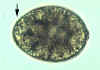 Figure 4A Figure 4A
Eggs of Diphyllobothrium latum. These eggs are oval or ellipsoidal, with at one end an operculum (arrows) that can be inconspicuous
(right). At the opposite (abopercular) end is a small knob that can be barely discernible
(left). The eggs are passed in the stool unembryonated. Size range: 58 to 76 µm by 40 to 51 µm.
CDC. Image A contributed by Georgia Division of Public Health
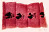 Figure 4B
Gravid proglottids of Diphyllobothrium
latum. CDC/Dr. Mae Melvin
 Figure
4C Figure
4C
Proglottids of Diphyllobothrium latum. The species characteristics are: the proglottid is broader than it is long; size 2 to 4 mm long by 10 to 12 mm wide; uterus coiled in rosette appearance; genital pore at the center of the
proglottid.
CDC
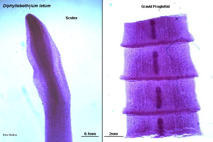
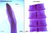 Figure 4E Figure 4E
Diphyllobothrium latum scolex and gravid proglottids
©
Dr Peter
Darben, Queensland University of Technology clinical parasitology
collection. Used with permission
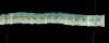 Figure 4D Figure 4D
Proglottids of Diphyllobothrium latum. These proglottids tend to be passed in strands of variable length in the stool. The proglottids tend to be broader than long.
CDC. Image contributed by Georgia Division of Public Health.
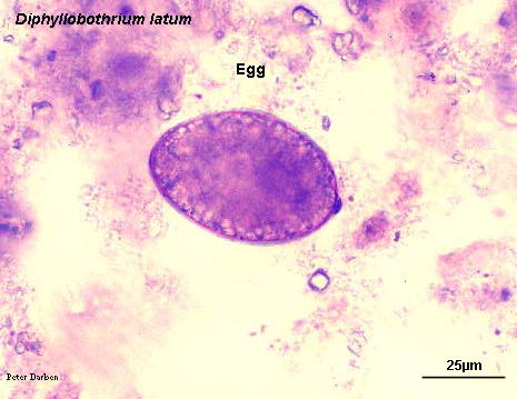
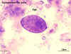 Figure 4F Figure 4F
Diphyllobothrium latum egg © Dr
Peter Darben, Queensland University of Technology clinical
parasitology collection. Used with permission
|
|
Figure
4
|
|
WEB RESOURCES
Hymenolepiasis
- CDC |
Hymenolepis nana (dwarf tapeworm)
This is a small tapeworm (20 x 0.7 mm)
which infects children. Rodents are the reservoir. Infection is by the oro-fecal
mode and, hence, cross infection and auto infection by eggs in feces in normal
(figure 6).
The worm develops from ingested eggs into an adult in the small intestine and
resides there for several weeks (figure 5). Light infections produce vague abdominal
disturbances but heavier infections may cause enteritis. Diagnosis is based on
finding eggs in the feces. Praziquantel is the drug of choice. Hygiene is the
best control.
|
|
|
 Figure 5
Eggs of Hymenolepis nana are immediately
infective when passed with the stool and cannot survive more than 10
days in the external environment
.
When eggs are ingested by an arthropod intermediate host
 (various species of beetles and fleas may serve as intermediate hosts),
they develop into cysticercoids, which can infect humans or rodents upon
ingestion
(various species of beetles and fleas may serve as intermediate hosts),
they develop into cysticercoids, which can infect humans or rodents upon
ingestion  and develop into
adults in the small intestine. A morphologically identical
variant, H. nana var. fraterna, infects rodents and uses
arthropods as intermediate hosts. When eggs are ingested
(in contaminated food or water or from hands contaminated with feces),
the oncospheres contained in the eggs are released. The
oncospheres (hexacanth larvae) penetrate the intestinal villus and
develop into cysticercoid larvae
.
Upon rupture of the villus, the cysticercoids return to the intestinal
lumen, evaginate their scoleces
,
attach to the intestinal mucosa and develop into adults that reside in
the ileal portion of the small intestine producing gravid proglottids and develop into
adults in the small intestine. A morphologically identical
variant, H. nana var. fraterna, infects rodents and uses
arthropods as intermediate hosts. When eggs are ingested
(in contaminated food or water or from hands contaminated with feces),
the oncospheres contained in the eggs are released. The
oncospheres (hexacanth larvae) penetrate the intestinal villus and
develop into cysticercoid larvae
.
Upon rupture of the villus, the cysticercoids return to the intestinal
lumen, evaginate their scoleces
,
attach to the intestinal mucosa and develop into adults that reside in
the ileal portion of the small intestine producing gravid proglottids
 .
Eggs are passed in the stool when released from proglottids through its
genital atrium or when proglottids disintegrate in the small intestine
.
An alternate mode of infection consists of internal autoinfection, where
the eggs release their hexacanth embryo, which penetrates the villus
continuing the infective cycle without passage through the external
environment .
The life span of adult worms is 4 to 6 weeks, but internal autoinfection
allows the infection to persist for years. CDC
DPDx
Parasite Image Library .
Eggs are passed in the stool when released from proglottids through its
genital atrium or when proglottids disintegrate in the small intestine
.
An alternate mode of infection consists of internal autoinfection, where
the eggs release their hexacanth embryo, which penetrates the villus
continuing the infective cycle without passage through the external
environment .
The life span of adult worms is 4 to 6 weeks, but internal autoinfection
allows the infection to persist for years. CDC
DPDx
Parasite Image Library
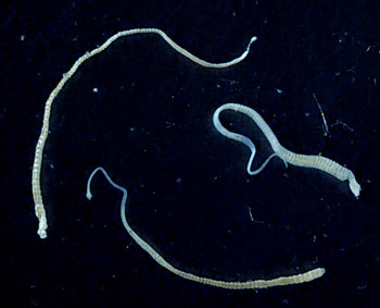
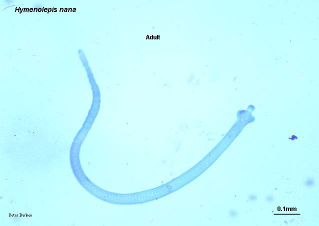
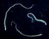 Figure 6A Figure 6A
Three adult Hymenolepis nana tapeworms. Each tapeworm (length: 15-40 mm) has a small, rounded scolex at the anterior end, and proglottids can be
distinguished at the posterior, wider end.
CDC. Image contributed by the Georgia Division of Public
Health.
 Figure 6B
Egg of Hymenolepis diminuta. These eggs are round or slightly oval, size 70 - 86 µm X 60 - 80 µm, with a striated outer membrane and a thin inner membrane. The space between the
membranes is smooth or faintly granular. The oncosphere has six hooks (of which at least four are visible at this level of focus).
CDC. Image contributed by Georgia Department of Public
Health.
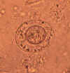 Figure 6C Figure 6C
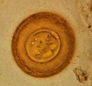
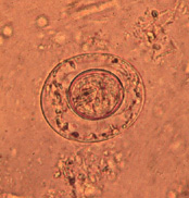
Egg of Hymenolepis nana. These eggs are oval or subspherical and smaller than those of H.
diminuta, their size being 40 - 60 µm X 30 - 50 µm. On the inner membrane are two poles, from which 4-8 polar filaments spread out between the two membranes. The oncosphere has six hooks (seen as dark lines at 8
o'clock). CDC. Image contributed by Georgia Department of Public
Health.
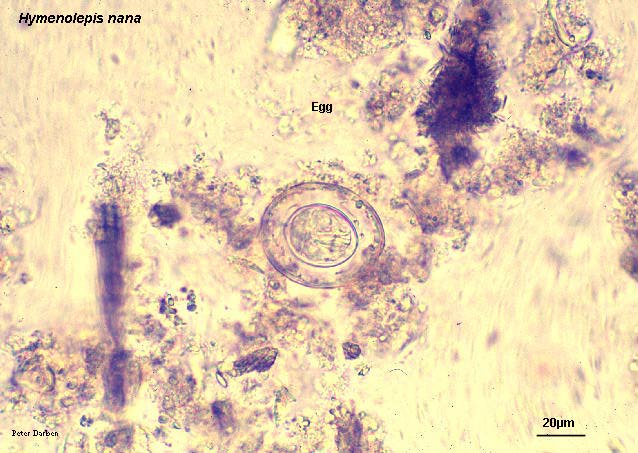
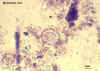 Figure 6D
Hymenolepis nana egg
© Dr
Peter Darben, Queensland University of Technology clinical
parasitology collection. Used with permission
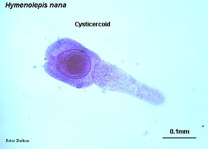
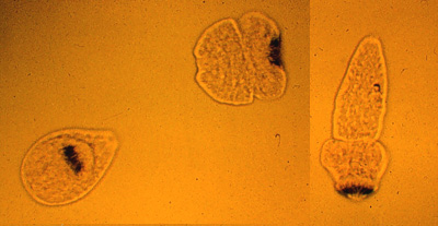
 Figure 6E
Hymenolepis nana cysticercoid
© Dr
Peter Darben, Queensland University of Technology clinical
parasitology collection. Used with permission
 Figure 6F
Hymenolepis nana adult
©
Dr
Peter Darben, Queensland University of Technology clinical
parasitology collection. Used with permission
|
|
|
| |
|
WEB RESOURCES
Echinococcosis
- CDC |
Echinococcosis (hydatid)
Echinococcus
granulosus and E. multilocularis are causative agents of hydatid cysts.
|
| |
Echinococcus granulosus
Epidemiology
The organism is common
in Asia, Australia, Eastern Africa, southern Spain, southern parts of South
America and northern parts of North America. The incidence of human infection
about 1 to 2 per 1000 population and may be higher in rural areas of affected regions.
Morphology
This is the smallest of
all tapeworms (3 to 9 mm long) with only 3 proglottids.
Life cycle
The adult worm lives in
domestic and wild carnivorous animals. Eggs, passed by infected animals, are
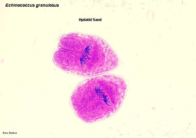
ingested by the grazing farm animals or man, localize in different organs and
develop into hydatid cysts containing many larvae (proto-scolices or hydatid
sand) (Figure 8). When other animals consume infected organs of these animals,
proto-scolices
escape the cyst, enter the small intestine and develop into adult worms (Figure
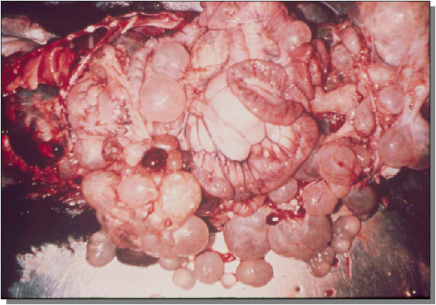
7). Echinococcus eggs, when swallowed by man, produce embryos that penetrate the
small intestine, enter the circulation and form cysts in liver, lung, bones, and
sometimes, brain. The cyst is round and measures 1 to 7 cm in diameter, although
it may grow to be 30 cm. The cyst consists of an outer anuclear hyaline cuticula
and an inner nucleated germinal layer containing clear yellow fluid. Daughter cysts
attach to the
germinal layer, although some cysts, known as brood
cysts, may have only larvae (hydatid sand). Man is a dead end host.
Symptoms
The symptoms, comparable to
those of a slowly growing tumor, depend upon the location of the cyst. Large
abdominal cysts produce increasing discomfort. Liver cysts cause obstructive
jaundice. Peribronchial cysts may produce pulmonary abscesses. Brain cysts produce
intracranial pressure and Jacksonian epilepsy. Kidney cysts cause renal
dysfunction. The contents of a cyst may produce anaphylactic responses.
Diagnosis
Clinical symptoms of a
slow-growing tumor accompanied by eosinophilia are suggestive. Intradermal (Casoni)
test with hydatid fluid is useful. Pulmonary cysts and calcified cysts can be
visualized using x-rays. Antibodies against hydatid fluid antigens have been
detected in a sizable population of infected individuals by ELISA or indirect
hemagglutination test.
Treatment and control
Treatment involves surgical removal of cyst or inactivation of hydatid sand
by injecting the cyst with 10% formalin and its removal within few (4-5)
minutes. Prazequantel has been shown to be effective in many cases. Albendazole,
in high doses, is an alternative. Preventive measures involve avoiding contact
with infected dogs and cats and elimination of their infection.
|
|
|
 Figure 7
The adult Echinococcus granulosus (3 to 6 mm long) (1) resides in the small bowel of the definitive hosts, dogs or other
canids. Gravid proglottids release eggs (2) that are passed in the feces. After ingestion by a suitable intermediate host (under natural conditions: sheep, goat, swine, cattle, horses, camel), the egg hatches in the small bowel and releases an oncosphere
(3) that penetrates the intestinal wall and migrates through the circulatory system into various organs, especially the liver and lungs. Inthese organs, the oncosphere develops into a cyst
(4) that enlarges gradually, producing protoscolices and daughter cysts that fill the cyst interior. The definitive host becomes infected by ingesting the cyst-containing organs of the infected intermediate host. After ingestion, the protoscolices
(5) evaginate, attach to the intestinal mucosa (6), and develop into adult stages
(1) in 32 to 80 days. The same life cycle occurs with E. multilocularis (1.2 to 3.7 mm), with the following differences: the definitive hosts are foxes, and to a lesser extent dogs, cats, coyotes and wolves; the intermediate host are small rodents; and larval growth (in the liver) remains indefinitely in the proliferative stage, resulting in invasion of the surrounding tissues. With E. vogeli (up to 5.6 mm long), the definitive hosts are bush dogs and dogs; the intermediate hosts are rodents; and the larval stage (in the liver, lungs and other organs) develops both externally and internally, resulting in multiple vesicles. E. oligarthrus (up to 2.9 mm long) has a life cycle that involves wild felids as definitive hosts and rodents as intermediate hosts. Humans become infected by ingesting eggs
(2), with resulting release of oncospheres (3) in the intestine and the development of cysts
(4) in various organs CDC DPDx
Parasite Image Library
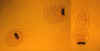 Figure
8A Figure
8A
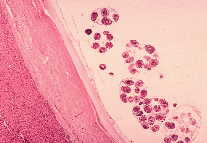
"Hydatid sand". Fluid aspirated from a hydatid cyst will shows multiple protoscolices (size approximately 100 µm), each of which has typical
hooklets. The protoscolices are normally invaginated (left), and evaginate (middle, then right) when put in saline.
CDC Image contributed by Georgia Division of Public Health
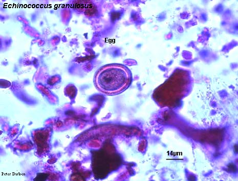
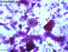 Figure 8B Figure 8B
Echinococcus granulosus egg
© Dr
Peter Darben, Queensland University of Technology clinical
parasitology collection. Used with permission
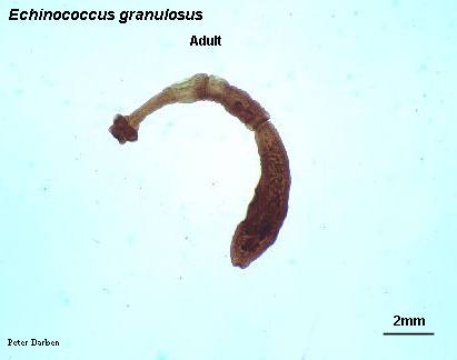
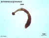 Figure 8C Figure 8C
Echinococcus granulosus adult
© Dr
Peter Darben, Queensland University of Technology clinical
parasitology collection. Used with permission
 Figure
8D Figure
8D
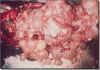
Echinococcus granulosus hydatid cysts in section of lung (H&E)
©
Dr Peter
Darben, Queensland University of Technology clinical parasitology
collection. Used with permission
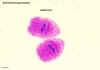 Figure 8E Figure 8E
Echinococcus granulosus hydatid sand
© Dr
Peter Darben, Queensland University of Technology clinical
parasitology collection. Used with permission
 Figure 8F Figure 8F
Histopathology of hydatid cyst. Echinococcus,
echinococcosis, CDC/Dr. Mae Melvin
 Figure
8G Figure
8G
Hydatid cysts
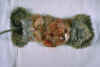 Figure 8F
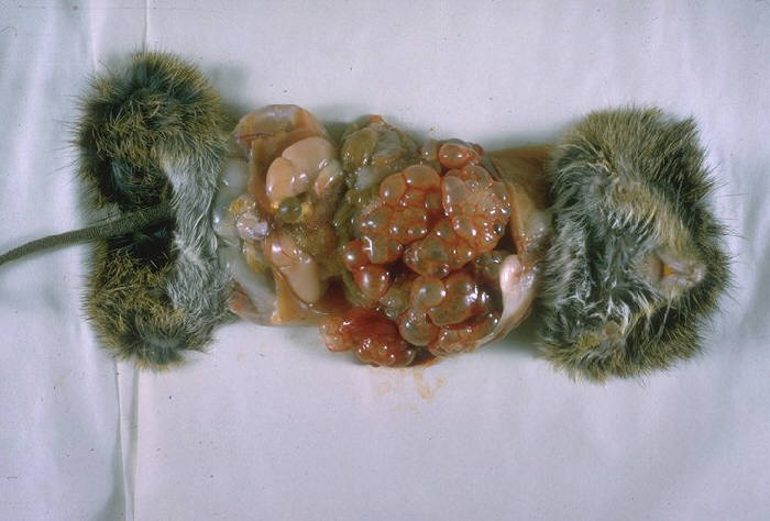
Gross pathology of cotton rat infected with Echinococcus
multilocularis. First E. locularis isolated in the United States proper.
CDC/Dr. I. Kagan
 Figure 8G Figure 8G
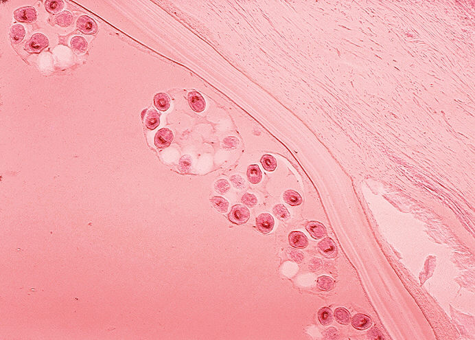
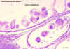
Histopathology of Echinococcus granulosus hydatid cyst in a sheep. Thick fibrous
pericyst, hyaline ectocyst, and brood capsules filled with
protoscolices are visible. CDC/Dr.Peter Schantz
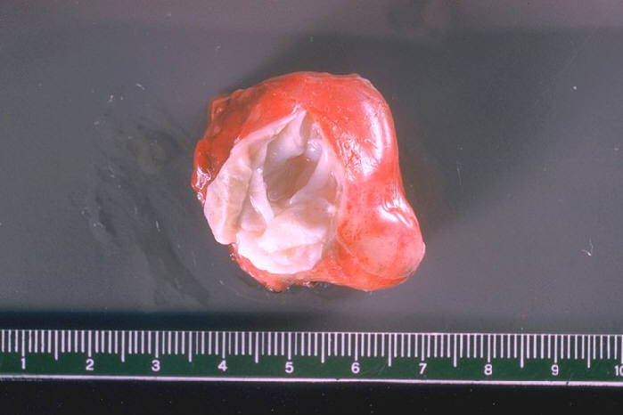
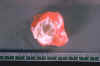 Figure 8H
Gross pathology of membrane and hydatid daughter cysts from human lung
CDC/Dr. I. Kagan
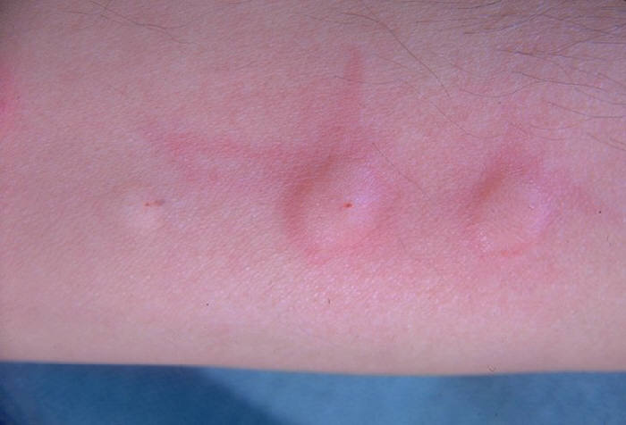
 Figure 8I
Man's arm showing positive skin test for hydatid disease
(echinococcosis) CDC/Dr. I. Kagan
|
|
Figure
8
|
| |
| |
| |
Echinococcus multilocularis
This is a tapeworm, similar to E.
granulosus, that also causes hydatid in northern parts of Asia and North America.
It has a very similar morphology and life cycle except that rodents are its
intermediate host. Humans, when infected with this worm, also develop hydatid
cysts which produce symptoms similar to those caused by E. granulosus. However,
the cysts are multilocular (many chambers). The organism is resistant to
praziquantel; high doses of Albendazole has some anti-parasitic effect. Surgery
is the means of removing the cyst. Rodent control is the means of prevention.
|
Summary |
|
Organism |
Transmission |
Symptoms |
Diagnosis |
Treatment |
| Tenia saginata
|
Cyst in beef |
Epigastric pain, vomiting, diarrhea |
Proglottids or eggs in stool or perianal area
|
Praziquantel |
| Tenia solium
|
Cyst in pork |
Epigastric pain, vomiting, diarrhea |
Proglottids or eggs in stool or perianal area
|
Praziquantel |
| T. solium Cysticercosis |
Oro-fecal |
Muscle pain and weakness, ocular and neurologic problems |
Roentgenography, anti-cysticercal antibody (EIA)
|
Praziquantel |
| D. latum
|
Cyst in fish |
Abdominal pain, loss of weight, anorexia, malnutrition and
B12 deficiency problems |
Proglottids or eggs in stool or perianal area |
Praziquantel |
| E. granulosus
|
Oro-fecal |
Large cysts produce various symptoms depending on the
location of the organism. |
Roentgenography,
anti-hydatid fluid antibody (EIA), Casoni
skin test |
Surgery, formalin injection and drainage, Praziquantel |
| E. multiloculoris |
Oro-fecal |
As above |
As above |
Surgery, Albendazole |
|
|
 Return to the Parasitology Section of Microbiology and Immunology On-line
This page last changed on
Tuesday, February 17, 2015
Page maintained by
Richard Hunt
|
 Figure 1
 Figure 3
 Figure 5
  Figure 7