Phylum: Microsporidia
Recently placed in the Kingdom Fungi, microsporidia are a large group of
intracellular parasites causing disease in animals including, especially,
invertebrates. They are emerging infectious agents because they can infect
patients living with HIV/AIDS or recipients of organ transplants and,
rarely, immune-normal persons.
Microsporidia reproduce with walled spores and infect host cells with a
structure called a polar filament. They must rely on the host to produce ATP
(Smith et al., 2017).
The next two fungal taxa were formerly in a Phylum, the Zygomycota. Now that
name and the term “zygomycete” have no strict taxonomic standing and are
informal terms.
Sub-Phylum: Entomophthoromycotina
Most are parasites of arthropods, some are saprobes and plant pathogens. Two genera contain zoopathogens-- Basidiobolus, and Conidiobolus. Disease occurs in subcutaneous tissues, nasal mucosae, and surrounding skin occurring mainly in tropical and sub-tropical Asia, Africa, and Latin America. The causative agents, however, are environmental fungi found worldwide.
Sub-Phylum: Mucoromycotina
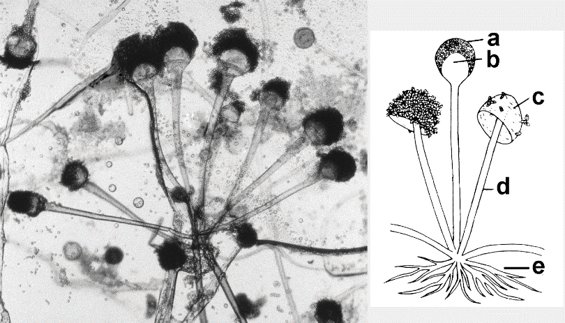
Mucorales are a fungal Order containing important pathogens, e.g.: Rhizopus, Rhizomucor, Mucor species among others (Figure 1). They are ubiquitous in the environment as soil saprobes, as plant pathogens, and cause postharvest rot of fruit. These opportunistic pathogens cause disease in immuno-compromised individuals, in ketoacidotic diabetics, and in burn patients. Mucormycosis includes cutaneous, mucocutaneous, and rhinocerebral disease; also renal infections, gastritis, and pulmonary infections. The infectious propagules of Mucorales are called spores and their presence in indoor environments implicates them as allergens.
Figure 1. Structural features of a mucormycete: Rhizopus oryzae
Left: A cluster of Rhizopus oryzae illustrates the “pin mold morphology.” The sporangiophores and sporangia are up to 1.5 mm in length, visible to the unaided eye. The sporangiophores, in a cluster of 8, may be simple or branched, rising from stolons opposite rhizoids. Columellae and the swollen basal part (apophysis) together are round or oval. Credit: Dr. Robert Simmons, Director, Biological Imaging Core Facility, Department of Biology, Georgia State University, Atlanta, GA.
inset: a. sporangium, b. columella, c. collapsed columella (after release of sporangiospores), d. sporangiophore, e. rhizoids. Credit: Kendrick, 1992, Dr. Bryce Kendrick and Focus Publishing R. Pullins Co.
The higher fungi are in the Sub-Kingdom: Dikarya
because in some part of their life cycle their cells contain two genetically
different haploid nuclei (one from each parent.) The dikaryotic state is
prolonged in basidiomycetes but short-lived in the ascomycetes.
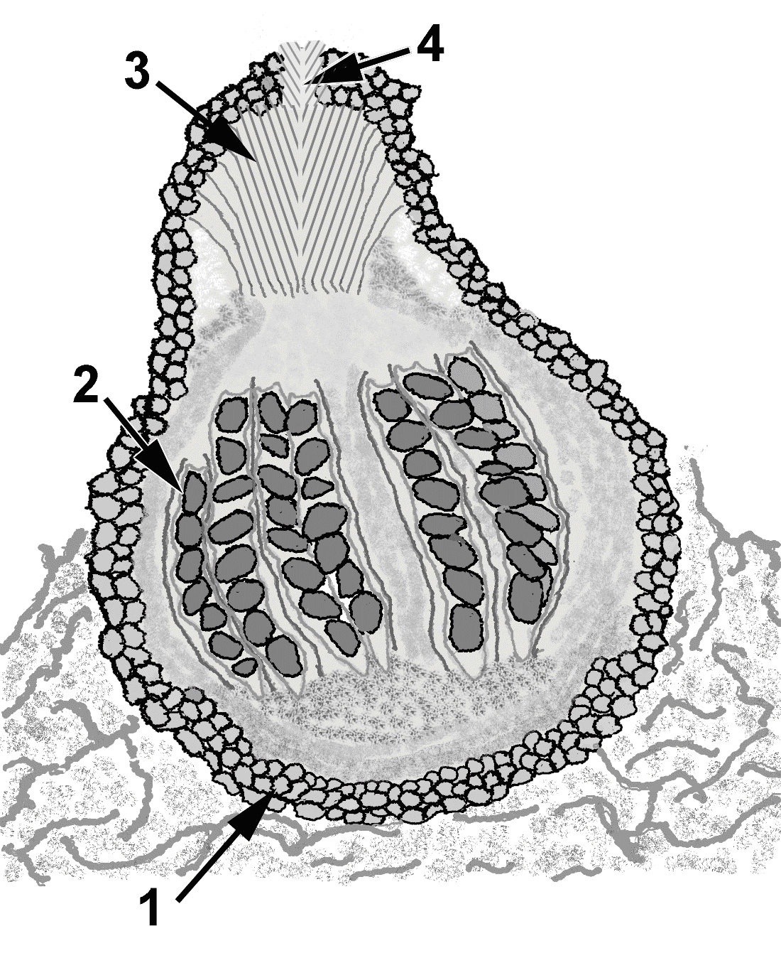
Phylum: Ascomycota.
The common theme for these “sac” fungi is sexual reproduction that occurs in
a sac (ascus) with the production of ascopspores (figure 2). The phylum
constitutes ~75% of all yeasts and molds (Taylor et al., 2006). Asexual
reproduction results in conidia which are infectious propagules, spread by
air, water, and by traumatic implantation via thorny plants and splinters.
The development, shape and size of conidia are important taxonomic features.
Examples of important pathogens classed here are: Candida species yeasts,
Aspergillus species molds, Pneumocystis; and the primary respiratory
pathogens: e.g.: Coccidioides species, Histoplasma capsulatum.
Figure 2
Drawing of a perithecium of an ascomycete, Sordaria species, in longitudinal section. 1. The peridium is the perithecium wall; 2. Asci and paraphyseal hyphae are the fertile layer of the ascoma: the hymenium; 3. Periphyseal hyphae provide a channel for escape of the ascospores through the 4. Pore or ostiole. Source: E. Reiss
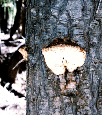
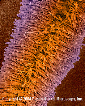
Phylum: Basidiomycota.
The name refers to the mode of sexual reproduction in a club-shaped basidium
with production of basidiospores (figures 3, A and B). Most basidiomycetes
are “mushrooms” with characteristic fruiting bodies, e.g.: hallucinogenic
and toxic Amanita muscaria (fly agaric). But in medical mycology the human
pathogenic basidiomycetes are, for the most part, yeasts! e.g.: Cryptococcus
neoformans, Malassezia species. C. neoformans is an opportunistic agent of
meningoencephalitis, notorious as one of the AIDS-defining opportunistic
infections. Malassezia species, on the other hand, are common commensals on
skin of mammals, including humans, causing superficial dermatomycosis.
A
B
Figure 3A. Example of a higher fungus that is a basidiomycete, a bracket fungus showing the basidiocarp (fruiting body).
Figure 3B. Lower surface showing generative hyphae. Reproductive spores are dispersed through pores in the surface of the brackets.
© Dr Arthur DiSalvo and © Dennis Kunkel Microscopy, Inc. Used with permission