| x | x | |||||
|
|
 |
|||||
| BACTERIOLOGY | IMMUNOLOGY | MYCOLOGY | PARASITOLOGY | VIROLOGY | ||
|
|
INFECTIOUS DISEASE |
|||||
|
|
||||||
|
Let us know what you think |
||||||
|
|
||||||
|
|
||||||
|
TEST YOUR KNOWLEDGE |
||||||
|
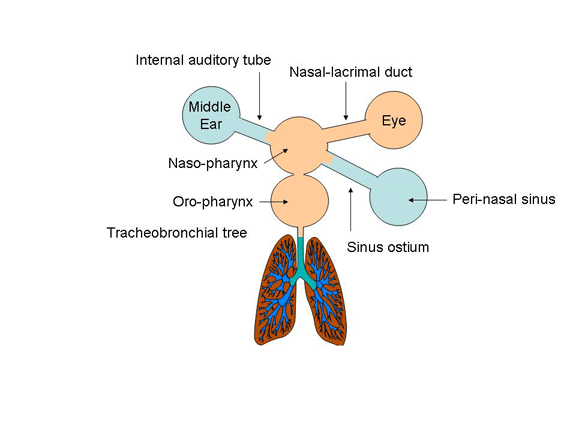
Upper respiratory tract infection (URI) causes at least of one-half of all symptomatic illness in the community, exacting huge tolls that can be measured as morbidity, absenteeism from school and work, direct health care costs, and overuse of antibiotics leading to the emergence of drug-resistant bacteria. This disease burden is largely explained by anatomy. The nose, mouth, and pharynx are exposed to circulating viruses and are normally colonized by large numbers of bacteria including potential pathogens such as S. aureus, S. pneumoniae, H. influenzae, and group A streptococci (figure 1). Mucosal injury caused by viral infection, allergy, or other factors compromises the mucociliary barriers designed to maintain sterility of the middle ears, paranasal sinuses, and lungs. Most URIs are self-limited but progression to life-threatening acute illness occurs and progression to chronic disease is common.
|
|||||
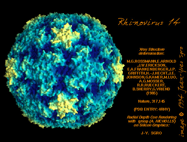 Figure 2 Human rhino virus © Dr J-Y Sgro, University of
Wisconsin. Used with permission
Figure 2 Human rhino virus © Dr J-Y Sgro, University of
Wisconsin. Used with permission
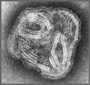
 Figure 4
Orthomyxovirus structure Lower image: CDC PHIL
|
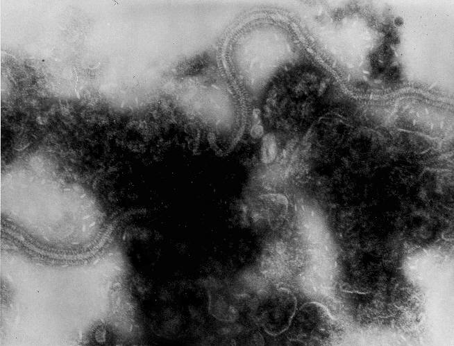
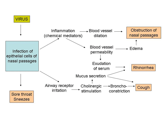
The Common Cold The common cold is as an acute, self-limited catarrhal (Latin catarrhus, to flow down) syndrome limited to the mucosal membranes of the upper respiratory tract. Recently, "viral rhinosinusitis" has been suggested as the technically correct term, since CT scanning shows abnormalities of the paranasal sinuses in most cases. However, "cold" is preferred for clinical use since "rhinosinusitis" suggests to patients the need for antibiotics. The common cold accounts for up to three-quarters of all illnesses in young infants and up to one-half of illnesses in adults. Elimination of the common cold would also eliminate in the United States each year an estimated 27 million office visits, 23 million days of work absenteeism, 26 million days of school absenteeism, and nearly $2 billion worth of over-the-counter remedies. Rhinoviruses (figure 2), of which there are more than 100 serotypes, cause an estimated 30% to 50% of colds. Coronaviruses account for perhaps 10% of cases. Respiratory syncytial virus (RSV) (figure 3) is an important cause of cold symptoms in young children and among the elderly. Influenza virus (an orthomyxovirus, figure 4) and parainfluenza virus (a paramyxovirus, figure 5) can cause colds, but more often cause lower respiratory tract infection or systemic symptoms. Adenoviruses, of which there are at least 47 antigenic types, cause 5% to 10% of colds. Although echoviruses and coxsackieviruses have been associated with colds, they more commonly cause an undifferentiated flu-like illness or distinctive syndromes such as aseptic meningitis or pharyngitis. Rhinoviruses and parainfluenza viruses cause outbreaks of cold symptoms during the fall and late spring; RSV, adenoviruses, and coronaviruses cause outbreaks during the winter and early spring. Viruses that remain to be discovered probably cause a substantial fraction of colds. Rhinoviruses are transmitted most efficiently by direct contact. Hand contact with the eyes and nose is very common in everyday life. Rhinoviruses remain viable on skin and also on objects (fomites) for at least 2 hours. Rhinoviruses can be recovered from hands in 40% to 90% of persons with colds and from up to 15% of objects near persons with colds. However, brief exposure such as a handshake or even being around an infected person for 36 hours causes transmission in less than 10% of subjects and in one study transmission was only 38% between spouses. Rhinoviruses can also be transmitted by aerosolization, for example, by being in a crowded room where people are sneezing. Kissing does not seem to be a common mode of transmission, probably because only about 10% of persons with colds have demonstrable virus in their saliva. Studies carried out in Antarctica dispel the popular idea that cold weather increases susceptibility to rhinovirus infection. The nasal epithelium of persons with colds is remarkably intact even when studied with the electron microscope. Symptoms are best explained by physiologic responses:
After an incubation period of 24 to 72 hours, most patients develop a sore or scratchy throat which is followed by nasal obstruction, rhinorrhea, and sneezing (figure 6). A green or yellow nasal discharge should not be construed as evidence of secondary bacterial infection (neutrophils cause yellow-green discoloration because of their natural myeloperoxidase activity). By the second and third day of the illness, rhinitis with nasal congestion replaces sore throat as the major complaint. By the fourth and fifth day, nasal symptoms have usually decreased but in about 30% of cases are replaced by cough or "chest cold".
|
|||||
|
Acute Bacterial Sinusitis Few common problems in primary care are as confusing as sinusitis. Acute bacterial sinusitis is vastly over diagnosed, but chronic sinusitis can be frustrating and disabling. The paranasal sinuses are accessible to direct examination only by sophisticated instruments. Adequate specimens for cultures can be obtained only by invasive procedures. Low-grade sinusitis is an intrinsic feature of the common cold. Clinicians must distinguish between self-limited viral rhinosinusitis and acute bacterial sinusitis, which usually calls for antibiotic therapy. Sinusitis is usually caused by obstruction of the ostia, as from edema, damage to ciliated epithelial cells, and/or increased volume or viscosity of the mucous secretions. The pathogenesis of acute sinusitis can be discussed in three complementary ways: anatomy, physiology, and microbiology.
Sinusitis usually begins with symptoms of the common cold such as runny nose, nasal obstruction, sore throat, cough, and the sensation of “pressure” or “tightness” in the face. The symptoms differ somewhat between children and adults. Children with sinusitis can have either of two presentations. The more common presentation consists of persistent cold symptoms that is, symptoms lasting more than 10 days. Children with persistent sinusitis seldom complain of headache or facial pain. Parents of young children often report malodorous breath. The less common presentation consists of severe cold symptoms―that is, cold symptoms that are accompanied by high fever (> 39° C) and purulent nasal discharge. Some of these children experience headaches usually located behind or around the eye, occasionally with periorbital edema. Adults with sinusitis, compared with children, tend to have more prominent facial pain, sometimes with local tenderness, swelling, and erythema. Pain patterns and other findings vary according to which sinuses are involved:
Low-grade fever is often present. Physical examination may reveal tenderness on percussion of the maxillary or frontal sinuses, and pinching the bridge of the nose may bring out tenderness if the ethmoid sinuses are involved. However, the sensitivity and specificity of these findings are unknown, and the clinical findings are often subtle. Acute bacterial sinusitis is often self-limited, but the frequency with which this condition resolves spontaneously is unknown. Direct sinus puncture for accurate diagnosis has not been carried out in placebo-controlled clinical trials. Serious complications occur often enough to justify close follow-up, especially in the clinically more severe cases. The most common complication is progression to chronic sinusitis (see below). Cavernous sinus thrombosis can result from ethmoid, frontal, or sphenoid sinusitis. Ethmoid sinusitis can also cause orbital cellulitis. Frontal sinusitis can also cause osteomyelitis of the frontal bone with swelling and edema of the forehead (Pott’s puffy tumor) and subdural empyema. Sphenoid sinusitis can be a medical emergency.
|
||||||
Chronic SinusitisWhen acute sinusitis fails to resolve and becomes chronic, cultures may reveal a variety of opportunistic pathogens including anaerobic bacteria. Some authorities feel that the problem is no longer mainly “infectious” but rather reflects permanent mucosal injury. It has been estimated that chronic sinusitis causes morbidity, measured as absenteeism from school, work, or social activities, of the same magnitude as heart disease and arthritis. The prevailing view holds chronic sinusitis to be a disorder of abnormal anatomy and physiology of the paranasal sinuses with one or more causes:
These processes lead to anatomic changes including obstruction of the infundibula and ostia, mucosal edema and scarring, bone hypertrophy, polypoid degeneration, and/or mucosal fibrosis, rendering the mucociliary clearance mechanism defunct. Numerous microorganisms can be isolated from patients with chronic sinusitis, but correlation between culture results and the disease process is often poor. Mixtures of aerobic and anaerobic bacteria are common. The general conclusion at this time is that in most patients, no single microorganism can be assigned a pathogenic role. In some patients, however, Pseudomonas aeruginosa or Staphylococcus aureus seems to be clearly pathogenic, and there are data suggesting roles for Haemophilus influenzae and Moraxella catarrhalis (in children). Patients with chronic sinusitis often have exacerbations analogous to the acute exacerbations of chronic obstructive lung disease. In these instances, especially in children, Streptococcus pneumoniae and Haemophilus influenzae may be important. Numerous bacteria including gram-negative rods have been isolated with patients with post-operative sinusitis. Some investigators believe that many patients with chronic sinusitis have allergic fungal sinusitis (discussed further below), the disease manifestations being caused by an immune response to extramucosal fungi. The typical history consists of nasal drainage, obstruction, and postnasal drip lasting for at least several months, often against a background of chronic “sinus trouble.” Patients often complain of headache or “sinus pain,” nocturnal cough, and bad breath. Loss of smell may also be present. Fever is unusual. A history of inhalant allergy is 4.5 times more common in patients with chronic sinusitis than in persons without chronic sinusitis. Although physical examination can reveal findings such as nasal septal deviation or mucosal changes, imaging studies are necessary to make a correct diagnosis. The coronal CT scan represents the current gold standard, but axial CT scans are often useful especially in children. Sinus endoscopy an also provide invaluable information. Chronic sinusitis may have a relapsing or remitting course, but, untreated, patients seldom become entirely free of symptoms related to the paranasal sinuses. Complications include remodeling of the facial bones, osteomyelitis, and―occasionally―invasive disease of the CNS caused by bacteria or fungi.
|
||||||
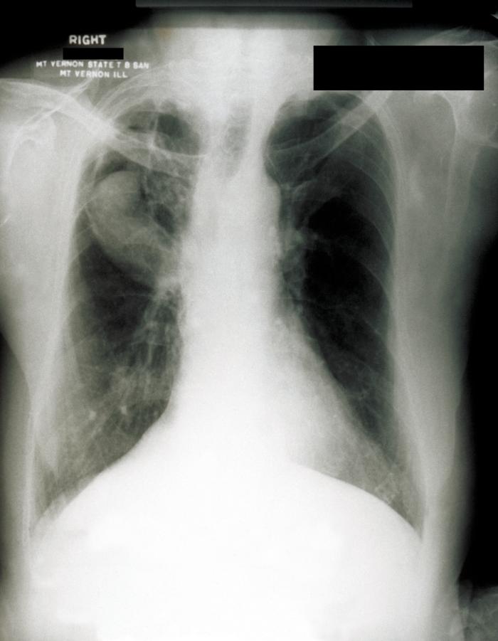 This chest radiograph shows probable aspergillosis with an aspergilloma,
or fungus ball in the upper lobe of the right lung. Lung diseases that
damage a lung can cause cavities that can leave a person more susceptible
to developing an aspergilloma, or fungus ball. The fungus can then begin
secreting toxic and allergic products, which may make the person feel ill.
CDC/M. Renz
This chest radiograph shows probable aspergillosis with an aspergilloma,
or fungus ball in the upper lobe of the right lung. Lung diseases that
damage a lung can cause cavities that can leave a person more susceptible
to developing an aspergilloma, or fungus ball. The fungus can then begin
secreting toxic and allergic products, which may make the person feel ill.
CDC/M. Renz
|
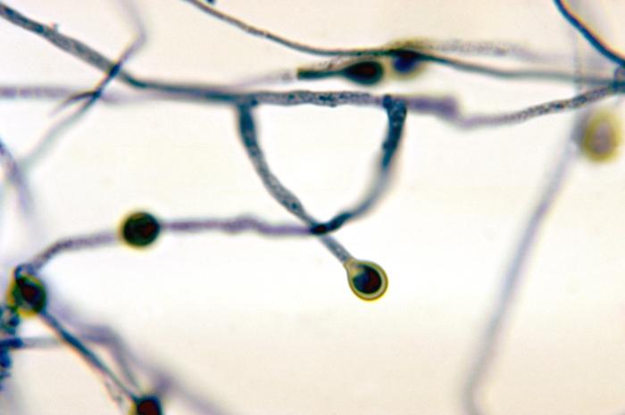
Fungal Sinusitis Fungal sinusitis is relatively uncommon, but should be considered in patients with chronic sinusitis because of its potentially serious complications. Aspergillus species are the most common causes of fungal sinusitis, and fungi of the order Mucorales are the most dangerous (rhinocerebral mucormycosis). Various widely distributed pigmented fungi that are collectively known as dematiaceous molds can cause a variety of syndromes that include life-threatening disease; examples of these organisms include Alternaria, Bipolaris, Cladosporium, Curvularia, and Exserohilum. Numerous other fungi sometimes cause sinusitis. Five syndromes are currently recognized.
|
|||||
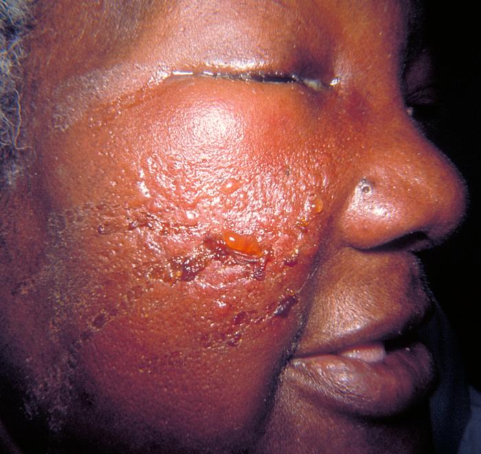 Erysipelas is a dermatologic condition, which involves the inoculation of the skin and subcutaneous tissue with streptococcal bacteria causing edema and bright red erythema of the affected areas. It is sometimes referred to as cellulitis. CDC |
Otitis Externa Otitis externa, a spectrum of conditions caused by infection, allergy, or primary skin disease, affects up to 10% of all people during their lifetimes. Necrotizing (malignant) otitis externa is a medical emergency seen usually in patients with diabetes mellitus or a compromised immune system or in patients who have had prior irradiation of the head. Otitis externa begins with breakdown in the cerumen barrier. Cerumen, although commonly considered a nuisance, protects against infection by (1) creating an acidic environment hostile to bacterial and fungal growth; (2) promoting a dry environment through its hydrophobic properties; and (3) trapping debris by its sticky nature. Excessive cleaning or scratching of the ear canal promotes breakdown of the cerumen barrier. Swimming notoriously predisposes to otitis externa, the main effect being promotion of a more alkaline pH, which in turn promotes bacterial growth. However, increased moisture of any origin leads to maceration of the skin and breakdown. Mechanical trauma from devices such as ear plugs (used for hearing conservation in many industries), headphones, hearing aids, and diving caps also predisposes to otitis externa. Pseudomonas aeruginosa and S. aureus are common causes of otitis externa. Group A streptococci and various gram-negative rods sometimes cause this condition, and anaerobic bacteria are involved in up to 25% of cases. About 10% of cases are caused by various fungi, with Aspergillus species being the most common followed by Candida species.
Diagnosis of otitis externa is nearly always made by the history and physical examination. Pressure on the tragus or pulling the auricle superiorly causes pain; the latter maneuver is a valuable diagnostic aid. Examination of the ear canal shows erythema and edema and, in severe cases, partial or complete occlusion of the canal. To exclude otitis media, one should demonstrate with pneumatic insufflation that the tympanic membrane is mobile. However, the tympanic membrane is often partially or totally obscured by edema in the ear canal. Although otitis externa is usually considered a self-limited condition, serious complications can occur. Perforation of the tympanic membrane can be caused by extension of the disease process or by misguided attempts by patients or health care providers to relieve the condition through mechanical manipulation. Other complications include stenosis of the ear canal, auricular cellulitis, or chondritis.
|
|||||
|
Acute Otitis Media The importance of otitis media in primary care cannot be overemphasized, especially in pediatrics practice, where it accounts for about 25% of all office visits, 50% of office visits for illness, and 40% of antibiotic prescriptions. By age 5, between 75% and 95% of children have had at least one episode of otitis media. This disease is responsible for some 25 million office visits each year, with annual health care costs estimated to be between 3 and 5 billion dollars. Although 80% of patients with otitis media are less than 15 years of age, more than one-fourth of all oral antibiotic prescriptions in the United States are written for this condition. Heavy prescribing of β-lactam antibiotics for otitis media is thought to be responsible in large measure for the decreasing drug susceptibility of S. pneumoniae strains. Acute otitis media is often preceded by viral URI that causes edema and obstruction of the eustachian tube, causing an ex vacuo serous transudate into the middle ear and paving the way for pathogenic bacteria. Children are predisposed because their eustachian tubes are shorter, wider, and straighter compared with those of adults. Appreciation of otitis media requires knowledge of the relationship of the eustachian tube to the nasopharynx, the middle ear, and the mastoid air cells. The eustachian tube normally serves to regulate pressure in the middle ear, to protect against nasopharyngeal sound pressure and secretions, and to afford a pathway for the drainage of secretions produced within the middle ear into the nasopharynx. The latter function requires an intact mucociliary system. Factors predisposing to eustachian tube dysfunction and otitis media include allergy, cleft palate, ciliary dysmotility, immunodeficiency, exposure to tobacco smoke, exposure to frequent upper respiratory tract infections (notoriously, in day care centers), early age of first infection, and race. Native Americans are markedly predisposed to otitis media for reasons that are unclear. Adults can be predisposed to otitis media on account of diabetes mellitus, cancer, immune deficiencies, and injection drug use. The adenoids have long been implicated in the pathogenesis of otitis media, for better or worse, since
The microbiology in otitis media is similar in adults and children when tympanocentesis is carried out. In about 40% of cases, culture of middle ear fluid fails to show a bacterial pathogen. This might reflect a viral etiology or sterile inflammation. Respiratory syncytial virus has been found relatively commonly, with a special tendency to cause the disease in children. Influenza viruses and parainfluenza viruses also predispose to otitis media. Streptococcus pneumoniae causes about 30% to 40% of cases, and the prevalence of strains with reduced susceptibility to penicillin is increasing. H. influenzae causes between 20% and 30% of cases, and M. catarrhalis between 10% and 15% of cases, especially in children. Group A streptococci cause less than 5% of cases but can cause up to 10% during the winter months. Acute otitis media in children usually presents with rapid onset of otalgia, fever, and/or irritability. Otalgia in young infants is manifest by pulling on the ear. Young children can also have anorexia, loose stools, and vomiting. Otalgia tends to be the major symptom in adults. A minority of patients experience spontaneous perforation of the tympanic membrane. The tympanic membrane is abnormal, often bulging, with loss of the usual landmarks. Erythema of the tympanic membrane alone is not diagnostic; it can be caused, for example, by crying. Purulent fluid is sometimes seen behind the tympanic membrane. The key procedures are pneumatic otoscopy and, when indicated, tympanocentesis. Pneumatic otoscopy, which is done by gently squeezing and then releasing a rubber bulb attached to the otoscope, provides information about the mobility of the tympanic membrane. Tympanocentesis provides fluid for culture, which is becoming more important due to the emergence of drug-resistant bacteria. Cultures of the nasopharynx correlate poorly with cultures of fluid obtained by tympanocentesis, and are therefore of limited usefulness. Acute otitis media must be distinguished from otitis media with effusion (serous otitis media). The latter consists of an asymptomatic or hyposymptomatic middle ear effusion, which can be acute (less than 3 weeks), subacute (3 weeks to 3 months), or chronic. Although hearing loss is frequently present in both acute otitis media and otitis media with effusion, patients with otitis media and effusion lack systemic signs and symptoms such as otalgia and fever. The natural history of untreated otitis media continues to prompt debate whether most cases should be treated with antibiotics. A meta-analysis of the literature based on data obtained from 5400 children in 33 studies indicates that 81% percent of children have spontaneous resolution. However, it is difficult to predict on clinical grounds whether an individual patients disease will resolve spontaneously, and the current consensus opinion in the United States is that all patients should be treated.
|
||||||
|
Chronic suppurative otitis media and mastoiditis Chronic suppurative otitis media is a complication of acute otitis media, usually occurring when there is a defect in the tympanic membrane, such as a "central" perforation or a tympanostomy tube. It is accompanied by purulent discharge (otorrhea). Mastoiditis is invariably present. The associated bacteria seem to vary depending on whether an infected cholesteatoma is present. Cholesteatoma is often associated with anaerobic bacteria and “skin flora” microorganisms, and the otorrhea often has a foul odor. When cholesteatoma is not present, gram-negative rods including Pseudomonas aeruginosa and E. coli are often found. In the pre-antibiotic era, mastoiditis was often a dramatic and severe illness with retroauricular inflammation and serious intracranial complications. Today, mastoiditis is more typically an indolent, low-grade, often painless infection of the temporal bone that tends to be clinically silent ("masked mastoiditis") unless a complication such as brain abscess develops. Patients at high risk of complications include newborn infants, persons with diabetes mellitus, the elderly, and the immunocompromised. Spontaneous resolution is rare, if it occurs at all. Local complications of chronic suppurative otitis media and mastoiditis include bone destruction, subperiosteal abscess, facial paralysis, labyrinthitis, and petrositis. Intracranial complications include brain abscess, subdural abscess, epidural abscess, septic thrombosis of the lateral sinus, meningitis, and hydrocephalus. Patients with chronic suppurative otitis media and/or mastoiditis should be referred to an otolaryngologist, as effective treatment usually requires surgical intervention.
|
||||||
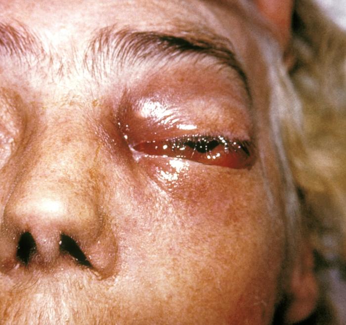
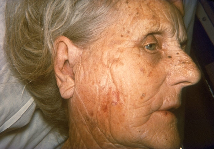 Figure
Staphylococcal parotitis, face of elderly woman. CDC |
Acute suppurative parotitis Acute suppurative parotitis, an uncommon condition, usually results from decreased salivary flow, allowing retrograde ascent of bacteria from Stenson’s duct. It can also result from ductal obstruction from mucus or fibrinous plugs, tumors, or foreign bodies). The clinical signs and symptoms include an acutely swollen, indurated check with fever and pain. Pus can be expressed from Stenson’s duct. More than 80% of cases are caused by Staphylococcus aureus. Most patients are elderly, debilitated, and dehydrated. Mortality is high.
|
|||||
|
Acute pharyngitis Acute pharyngitis (sore throat) is one of the most common problems encountered in clinical practice. Viruses cause most cases as part of the common cold. However, about 15% of cases, and up to 50% of cases in children during some periods, are caused by group A β-hemolytic streptococci (S. pyogenes). Although usually self-limited, streptococcal pharyngitis demands respect as a cause of acute rheumatic fever and―less commonly―major suppurative complications, acute glomerulonephritis, and even the streptococcal toxic-shock syndrome. The clinician’s task is to determine in a cost-effective manner which patients need treatment and which do not. Acute pharyngitis has many known etiologies, and pathogens remain to be discovered for an estimated 30% of cases. Viral infections cause sore throat, it is thought, by generating bradykinin and lysyl bradykinin, which stimulate nerve endings. Group A streptococci and certain other pathogens including some of the respiratory viruses cause pain by invading the mucosa. Group A streptococci are carried in the human nasopharynx and transmitted from person to person usually by direct contact with saliva or nasal secretions. Acquisition is greatest in school-aged children, suggesting the gradual development of immunity over time. Children also serve as a reservoir for spread among family members. Asymptomatic pharyngeal carriage of group A streptococci is relatively common, and the factors that cause some persons to develop acute pharyngitis and other complications are poorly understood. Group C and group G streptococci cause a pharyngitis syndrome clinically indistinguishable from that caused by group A streptococci, sometimes recognized as outbreaks related to a common food source. Group C streptococci (S. dysgalactiae subspecies equisimilis) appear to be a frequent cause of pharyngitis in college-aged students. Pharyngitis due to group A streptococci occurs most frequently in children between 5 and 15 years of age, usually during the winter and early spring. In its severe form the disease starts abruptly with fever, sore throat, and odynophagia. Chills, headache, and abdominal pain are sometimes present. Examination reveals diffuse erythema of the pharynx and tonsils accompanied by a patchy, purulent tonsillar and pharyngeal exudate, hypertrophy of the lymphoid nodules in the posterior pharyngeal mucosa, and tender cervical lymphadenopathy. Occasional strains of S. pyogenes elaborate the erythrogenic toxin of scarlet fever, resulting in a striking rash and "red strawberry tongue" with enlargement of the papillae. Rhinorrhea and cough are usually not present, but may occur. However, these dramatic manifestations are absent in many, perhaps most cases of streptococcal pharyngitis. Because the features of group A streptococcal pharyngitis blend imperceptibly with those of other causes of sore throat, numerous students of the disease have concluded that the diagnosis must be secured by laboratory methods prior to definitive treatment (see below). Other syndromes include the following:
Miscellaneous causes of sore throat include juvenile rheumatoid arthritis, systemic lupus erythematosus, bullous pemphigoid, Behçet’s disease, paraquat ingestion, and drug reactions.
|
||||||
|
Acute Laryngitis Acute laryngitis is extremely common, usually occurring as part of upper respiratory tract infection. Treatment is symptomatic, but prolonged hoarseness mandates the search for other etiologies. Acute laryngitis is most often caused by respiratory viruses, but vocal abuse or gastroesophageal reflux must also be considered. Parainfluenza viruses are the usual cause in patients between ages 5 and 15. Hoarseness complicates up to 29% of rhinovirus infections, 35% of influenza virus and adenovirus infections, and 63% of coronavirus infections according to various studies. Hoarseness can also complicate acute streptococcal pharyngitis. H. influenzae and Moraxella catarrhalis are often isolated, but their pathogenic roles are unclear. Rarely, fungi such as Candida species, Cryptococcus neoformans and Coccidioides immitis can cause laryngitis. Uncommonly, laryngitis can also be caused by tuberculosis and blastomycosis. Acute laryngitis presents as hoarseness. Speech and/or swallowing may be painful. The voice is hoarse, harsh, broken, or nearly absent. There are often concomitant symptoms of common cold including sore throat. Diagnosis of acute laryngitis is made on clinical grounds, typically by the history. Direct or indirect laryngoscopy shows erythema and edema of the vocal folds, sometimes with submucosal bruising or micro-hemorrhages (if the patient continues major voice use or has a bad cough). Viral cultures and special studies are seldom indicated.
|
||||||
|
Odontogenic Infections Odontogenic infections, the most common infections of the oral cavity, begin in and around the teeth and can spread to cause life-threatening local or systemic complications. The normal oral cavity contains dense masses of bacteria, of which more than 80% or more are anaerobic. Viridans streptococci (such as S. mitis, S. sanguis, S. salivarius, and S. mutans) preferentially colonize one or another anatomic site. Streptococcus mutans is the major cause of dental caries (cavities). As is well known, poor oral hygiene facilitates development of dental plaque, composed mainly of anaerobic bacteria. Diet high in simple sugars and carbohydrates predisposes to plaque. Periodontal disease, on the other hand, is unrelated to diet but is associated with poor oral hygiene, increasing age, and with various congenital immunodeficiency diseases and juvenile diabetes mellitus. Gingivitis and periodontitis are caused mainly by anaerobic bacteria. Once established, suppuration arising in and around the teeth can spread along fascial planes, invade bone, or enter the bloodstream. Deeper infections requiring surgical drainage, such as periapical abscesses and deep fascial space infections, are usually associated with numerous anaerobic bacterial species. Odontogenic infections present as one of several syndromes:
Diagnosis of dentoalveolar infections, gingivitis, and periodontal infections is based upon symptoms, examination, and, when indicated, dental x-rays. Dental x-rays are especially valuable for visualizing periapical abscess and acute alveolar abscess.
|
||||||
|
Mouth Ulcers Careful examination of the oral cavity often provides evidence of important systemic and local disease. More commonly, primary care clinicians evaluate patients with self-limited diseases such as stomatitis due to the herpes simplex viruses and aphthous stomatitis (canker sores). Here we will briefly review these latter conditions and their differential diagnosis. Herpes simplex viruses 1 and 2 cause fever blisters
and, less commonly, lesions elsewhere in the mouth including the palate. The
cause of aphthous stomatitis is unknown. Current opinion favors an
immunopathogenesis involving T-cell immunity, possibly a delayed-type
hypersensitivity reaction to an antigen residing within the epithelium. Some
cases of aphthous stomatitis have been attributed to drugs. Stress, smoking,
hormonal factors, and food allergy have also been invoked, but the evidence is
unconvincing.
|
||||||
|
|
||||||